*Corresponding Author:
Mark J. O’Connor, AstraZeneca, Cambridge, United Kingdom.
Citation:
Lenka Oplustil O’Connor, Anderson Wang, Claire Sadler, Jennifer Barnes, Rajesh Odedra, Aaron Smith, Gareth Hughes, Alan Lau, Andres Tellez, Scott Eliasof, Elaine Cadogan, Mark J. O’Connor (2026), Gap Scheduling of a PARP Inhibitor and Nanoparticle TOP1 Agent Combination Avoids Synergistic Bone Marrow Toxicity; J. Clinical Cancer and Oncology, 3(4): DOI: 10.64585/3065-3320-3-4-09.
Copyright
:
2026 Mark J. O’Connor. This open-access article is distributed under the terms of The Creative Commons Attribution License, which permits unrestricted use, distribution, and reproduction in any medium, provided the original author and source are credited.
Received:12 June 2026
| Accepted:20 June 2026
| Published:24 July 2026
Keywords:
gap schedule, PARP inhibitor, TOP1 inhibitor, tumor-targeted delivery, Camptothecin
Although combinations of DNA damage response inhibitors (DDRi) and DNA-damaging chemotherapy enhance cytotoxicity in cell-based systems, clinical success has been limited by overlapping bone marrow toxicities. Here, we show that the tumour-targeted nanoparticle camptothecin CRLX101, when administered concurrently with DDRi, enhances anti-tumour efficacy but also increases bone marrow toxicity in preclinical models. In NCI-H417 small cell lung cancer cells, PARPi, ATRi, and ATMi all potentiated CRLX101 activity, with the PARPi olaparib reducing the CRLX101 growth inhibition (GI50) by 3.2-fold, ATR inhibition by 3.6-fold, and ATM inhibition by 7.8-fold. Following assessment of the most promising combinations in a rat bone marrow safety model, we identified olaparib as the best tolerated combination with CRLX101. However, concurrent dosing of CRLX101 with olaparib still increased hematological toxicity relative to either single agent alone. Using rat bone marrow progenitor cells as biomarkers and leveraging the differential repair kinetics of CRLX101 induced DNA damage in tumour and bone marrow, we identified a delayedor gap schedule of olapariband CRLX101 in which separation of dosing by 48 hours reduced the hematological impact of the combination while maintaining enhanced anti-tumour efficacy over single agents in xenograft models. Extended olaparib dosing after CRLX101 further improved tumour control, consistent with prolonged tumour retention of CRLX101. A clinical trial has been designed using the gap schedule identified here, and this approach may provide a template for combining DDR inhibitors with tumour-targeted DNA-damaging chemotherapy in a clinically translatable manner.
INTRODUCTION
DNA damaging chemotherapy has been at the centre of cancer therapy for over seven decades (Cheung-Ong et al., 2013; DeVita and Chu, 2008). Underlining the preferential sensitivity of tumours compared to normal tissues are deficiencies in the DNA damage response (DDR), a collective term for the intra- and inter-cellular signaling events involved in the detection and repair of DNA damage (Ciccia and Elledge, 2010; Jackson and Bartek, 2009). In spite of the cancer-specific DDR deficiencies (Jackson and Bartek, 2009), the levels of exogenously generated DNA damage causedby systemic chemotherapies still results in significant and unwanted side effects. For this reason, there has been an attempt over the last twenty years or so, to identify specific inhibitors of the DDR in order to exploit the same cancer-specific DDR deficiencies (Curtin, 2012; O'Connor, 2015). In the case of inhibitors of poly (ADP-ribose) polymerase (PARP), this has led to the first approved DDR-based medicines, where PARP inhibitors (PARPi) have demonstrated monotherapy activity (Coleman et al., 2017; de Bono et al., 2020; Golan et al., 2019; Ledermann et al., 2014; Litton et al., 2018; Mirza et al., 2016; Robson et al., 2017) through a mechanism described as synthetic lethality(Fong et al., 2009; Lord and Ashworth, 2017). However, the potential for PARP inhibitors goes well beyondmonotherapy in HRR-deficient cancers and includes the potentiation of DNA damaging chemotherapy in a broader range of tumours (Pilie et al., 2019). Moreover, in addition to inhibitors of PARP, there are now several other DDR targeted agents being developed in the clinic (O'Connor, 2015), including those agents that inhibit WEE1 (Hirai et al., 2009), ATR (Min et al., 2017; Reaper et al., 2011; Wengner et al., 2020), CHK1 (Angius et al., 2020), ATM (Durant et al., 2018; Riches et al., 2020), DNA-PK (Fok et al., 2019; Wise et al., 2019) and Aurora Kinase B (Ashton et al., 2016).
While there are no shortagesof preclinical exampleswhere DDR inhibitors (DDRi) enhance the anti-tumour activityof chemotherapies, in most cases, poor tolerability for these combinations is underestimated becauseof the use of sub-optimal preclinical models. For example, mouse tumour efficacy models will not accurately reflect the potential impact on bone marrow since mice preferentially use different (more efficient but error-prone) DDR pathways in bone marrow stem cells than either rats or humans (Lane and Scadden, 2010; O'Connor et al., 2016).For this reason, rats represent a better preclinical safety model than mice to assess bone marrow toxicity. When DDRi are combined with chemotherapy, the experience from the clinic is that the overlapping toxicities with DDRi present a significant challenge and currently there have been few successful trials demonstrating efficacy coupled with acceptable tolerability. This point is highlighted by the fact that currently there isn’t a single approval for a targeted DDR agent in combination with chemotherapy.
One approach to improve the therapeutic window of chemotherapy is to target the DNA damaging agent to the tumour and/or avoid the bone marrow compartment. Approaches to achieve this currently in development include re-formulation (e.g. through liposomal or nanoparticle approaches (Wang et al., 2012)) or conjugation of the chemotherapy war-headto an antibody that preferentially targets a tumour surface marker (antibody-drug-conjugates or ADCs; (Beck et al., 2017; Thomas et al., 2016)). However, even tumour-targeted delivery may not fully prevent overlapping toxicity when combined concurrently with DDR inhibitors.
Here, we investigated whether a rationally designed dosing schedule could widen the therapeutic window of a tumour-targeted topoisomerase I inhibitor when combined with DDR inhibition. We evaluated CRLX101, a nanoparticle camptothecin formulation CRLX101(Weiss et al., 2013), which we chose for combination studies based on its monotherapy activity, preferential accumulation in tumors, and favorable activity compared to conventional topoisomerase I inhibitors (TOP1i) (Clark et al., 2016; Pham et al., 2015; Weiss et al., 2013). For the DDRi combinations, we assessed the PARPi olaparib (Menear et al., 2008), WEE1i adavosertib/AZD1775 (Hirai et al., 2009), ATRi ceralasertib/AZD6738 (Foote et al., 2018), ATMi AZD0156 (Riches et al., 2020), DNA-PKi AZ’6119, a precursor of AZD7648 (Fok et al., 2019), and Aurora kinase Binhibitor AZD1152 (Wilkinson et al., 2007). After triaging combinations in vitro to identifythose with enhancedanti-tumour activity, we evaluated the most promising regimens in a rat bone marrowsafety model and in mouse xenograft efficacy studies. Our objective was to determine whether differential tumour and bone marrow damage-repair kineticscould be exploited to develop a gap schedule that reduces hematological toxicity without compromising efficacy, therebyproviding a clinically translatable starting point for combination testing.
METHODS
Cell culture and chemicals
All cell lines were grown at 37°C in humidified incubator with 5% CO2 and maintained in phenol-free Dulbecco Modified Eagle Medium (Gibco) supplemented with 10 percentage fetal bovine serum and 2mM GlutaMax (Gibco). AZD2281 (olaparib; PARPi), AZD1775 (adavosertib; WEE1i), AZD0156 (ATMi), AZD6738 (ceralasertib; ATRi), AZ’6119
(DNA-PKi), and AZ1152 (Aurora B kinase inhibitor) were synthesized internally at AstraZeneca. All compounds were dissolved in DMSO at stock concentration of 10 mM.
Cell growth inhibition assay
SCLC NCI-H417 cells were seeded in 96-well plates and allowed to adhere overnight before treatment with DMSO, CRLX101, or CRLX101 in combination with the indicated DDR inhibitors. For CRLX101 single-agent assessment, cells were exposed for 48h before drug washout. DDR inhibitors were added concurrently with CRLX101 and replenished after 48h. Growth inhibition (GI50) values were determined on day 7 using the MTT assay,in which tetrazolium MTT substrate (Sigma) was added to a final concentration of 0.25 mg/mL. Following 6–8h incubation, formazan crystalproducts were solubilized in 5% SDS and 0.005 M HCl (final concentrations). Optical density was read at 570nm, and absorbance values were normalized to the DMSO control. Relative absorbance values were analysed in GraphPad Prism to derive GI50 values.
Cell line immunoblotting and antibodies
Whole cell lysates were prepared by lysing cell pellets directly in 2x Laemmlisample buffer (4% SDS, 20% glycerol, 125 mM Tris-HCl pH 6.8) and vortexing samples at highest speed for 20 seconds. Following protein concentration measurement using DC Protein Assay (Bio-Rad), protein samples were supplemented with sample reducing agents at 1x (Invitrogen) and 0.01% bromophenol blue final concentration. Samples were boiled at 95°C for 5 minutes before proteins were separated on NuPAGE 4-12% Bis-Tris protein gels (Invitrogen) by SDS-PAGE.Following protein transferonto nitrocellulose membranes, immunoblotting analyses were performed using antibodies listed below.
Target
Company
Catalogue No
Origin
Dilution
ATM pS1981
Abcam
ab81292
rabbit
1/500
CDK1 pY15
Cell Signaling Technology
9111
rabbit
1/1000
CHK1 pS345
Cell Signaling Technology
2348
rabbit
1/500
Histone H3 pS10
Cell Signaling Technology
9701
rabbit
1/1000
γH2AX
Cell Signaling Technology
2577
rabbit
1/1000
RPA32 pS4/S8
Bethyl Laboratories
A300-245A
rabbit
1/1000
Vinculin
Sigma-Aldrich
V9131
mouse
1/5000
Cell cycle analysisby flow cytometry
Cells treated with indicated compounds were harvested and fixed in ice-cold70% ethanol overnightat -20 °C. Fixed cells were then washed with PBS before incubated in PBS containing 100 µg/ml RNase A (Thermofisher) and 50 µg/ml propidium iodide (Sigma) for 30 minutes at 37 °C. FACSCalibur (BD Biosciences) was employed to analyze samples, and data were plotted using FlowJo software.
In vivo efficacy and pharmacodynamic studies
All in vivo studies were performed within AstraZeneca in the United Kingdom, and study protocols were reviewed and approved by the Home Office. All studies were conducted in accordance with the Animal Scientific Procedures Act 1986 (ASPA) and AstraZeneca Global Bioethics policy. Data were reported in line with the Animal Research: Reporting In Vivo (ARRIVE) guidelines (Kilkenny et al., 2010). For efficacy studies, human small cell lung cancer cells (NCI-H417) were implanted subcutaneously in athymic Fox1-nu mice. Animals were dosed with individual agents or combinations of CRLX101 (once weekly) with or without DDR inhibitors. Animals were randomized into vehicle and treatment groups based on a mean tumour volume of approximately 0.1–0.2cm3. Treatment groups received CRLX101 alone at either 4mg/kg or 5mg/kgonce weekly, while DDR inhibitor daily oral dosing was 100mg/kg for olaparib, 120mg/kg for AZD1775, and 5mg/kg for AZD0156. Animals were monitored throughout the study according to institutional welfare guidance, and tumour and tissue sampling time points were selected to capture both early and sustained pharmacodynamic effects following CRLX101 exposure. Statistical significance was evaluated using a one-tailed t test.
In vivo rat studies
RccHan:WIST rats (n = 7 per treatment group) were obtained from Harlan UK. Animals were approximately 11 weeks old at the start of dosing, were acclimatized for at least 1 week, and were group-housed at up to 5 per cage. Water and RM1 (E) SQC pelleted diet (Special Diet Services Ltd, England) were provided ad libitum. Environmental enrichment included nesting material and polycarbonate tunnels. CRLX101 was formulated as a 2mg/mL PBS nanosuspension and dilutedto the required concentration in PBS on the day of use. Animals receiving CRLX101 were given a single intravenous administration on day 1. Olaparib was formulated in dimethyl sulphoxide diluted 1:10 with 10% hydroxypropyl-β-cyclodextrin in phosphate-buffered saline (pH 7.4) and administered once daily by oral gavage. When AZD2281 and CRLX101 were dosed on the same day, the oral olaparib dose was given approximately 1h after the intravenous CRLX101 dose. Topotecan was supplied by Accord Healthcare Limited, formulated in 0.9% saline, and administered daily by oral gavage. All animals were euthanized by administration of halothane.
Flow cytometry analysisof rat bone marrow cells
Rat femurs were removed at necropsy and both ends were trimmed. Bone marrow cells were immediately flushedout with 3 ml PBS containing 50 percentage fetal calf serum (FCS). Cell suspension was syringed and filtered through 100 μm strainer, collected by centrifugation (300 g/ 7 min/ 4ºC) and washed once in HBSS containing 2 percentage FCS and 10 mM HEPES (staining buffer). Total cell count of isolated cells was determined by automated cell counter (Countess, Invitrogen). Cell concentration was adjusted to 1x10⁷ cells/ ml in staining buffer and processed for antibody staining. CD71 and CD45 cocktail: 100 μl cell suspension wasresuspended in 100 μl stainingbuffer containing anti-rat CD71-FITC (dilution 1:10) and CD45-PE (1:10) from Serotec. Cells were incubated with antibodies for 30 minutes RT and washed twice in stainingbuffer. Cell pellet stained with CD71-CD45 antibodies was resuspended in 200 μl staining buffer. This was followed by additionof 10 μl of LDS-751 cell-permeant nuclear stain (Life Technologies). CD90.1 and Lineages cocktail: 1 ml of cell suspension was resuspended in 100 μl staining buffer containing anti-ratCD90.1-APC (dilution 1:100),CD6-FITC (1:100),CD3-FITC (1:100), CD11b-FITC (1:200), Granulocytes-FITC (1:200) from BD Pharmingen and CD45RC-FITC (1:100) purchased from Serotec. After final centrifugation, 1 ml of staining buffer was added to CD90.1-Lineages-stained cells. Cells were incubated in dark for 30 minutes prior to flow cytometry analysis. Data (at least 10,000 events) were acquired on FACS AriaII (BD). Analysis was performed in FlowJo software.
Immunohistochemistry (IHC) analysis of gH2AX in rat bone marrow and xenofraft tumor
Rat femurs or human tumour samples implanted in mice were removed at necropsy and fixed in formalin in situ for 48 hours prior to coring. Preserved tissue was processed into wax blocks, sectioned and stained by immunohistochemistry for the presence of gH2AX (Ventana™, Omin-UltraMap HRP, Doscovery XT Staining module; gH2AX CST 25777 @ 1:100). Sections were counter stained with haematoxylin. The slides providedfor analysis were scanned using the Aperio Scanscope, converted to .TIF images (x 10 magnifications), and analysed on the KS400 image analyser.
Haematology analysis of rat plasmasamples
Blood samples to be taken from the tail vein (0.4 mL into EDTA). Haematology analysis was performed on the same day using the Siemens Advia 2120i automated haematology analyser.
Statistical analysis
GI50 values from cell growth inhibition assays were calculated using GraphPad Prism by fittingnormalized dose-response curvesto the experimental data. For in vivo efficacy studies, tumour volume data were summarized over time, and statistical comparisons between treatment groups were performed using a one-tailed t test, as described in the study methods. A p value <0>
Data Availability Statement
The data supporting the findings of this study are contained within the manuscript and its supplementary materials. Additional underlying data may be made available by the corresponding author upon reasonable request, subject to institutional and confidentiality requirements.
RESULTS
DDR inhibitors differ in both the extent and mechanism of CRLX101 potentiation
CRLX101is a targeted topoisomerase I inhibitor (TOP1i)in clinical development based on a nanoparticle camptothecin formulation (Clark et al., 2016). Small cell lung cancer (SCLC) is a tumour type of interest for CRLX101 development because these tumours are responsive to TOP1 inhibitors, including the standard-of-care (SoC) therapeutic topotecan and remains an area of high unmet clinical need. To evaluate the benefit of combining CRLX101 with different DDR inhibitors, we chose to use the SCLC cell line model NCI-H417, because the responseto single agent CRLX101 had previously been characterized and importantly, the NCI-H417 cell line could be used to generate an in vivo xenograft model.
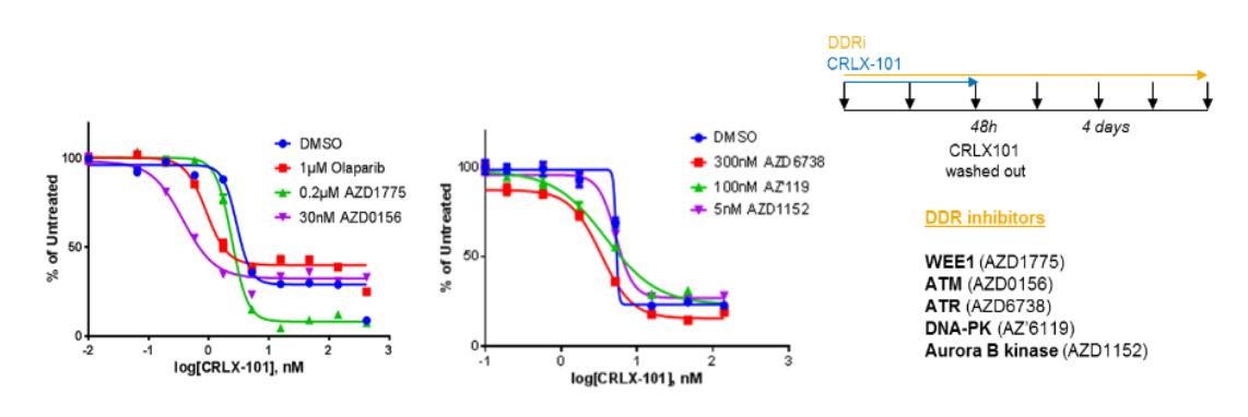
We treated the NCI-H417 cells with CRLX101 in combination with a PARP inhibitor (olaparib), a WEE1 inhibitor (AZD1775), an ATM inhibitor(AZD0156), an ATR inhibitor (AZD6738), a DNA-PK inhibitor (AZ’6119)and an Aurora kinase B inhibitor (AZD1152). Cells were exposed to CRLX101 for 48h, followed by drug wash-out. A fixed dose of DDR inhibitor (shownto provide greaterthan 95% target inhibition) was added concurrently to varying concentrations of CRLX101 on day 1 and replenished after 48h. Cell viability was determined at day 7 using an MTT assay (Figure 1A). The PARPi, ATRi and ATMi all significantly potentiated the activity of CRLX101, resulting in a decrease in the GI50 of CRLX101 by 3.2-fold, 3.6-fold and 7.8-fold respectively (Figure 1A).
Figure 1(A): Potentiation of CRLX101 by different DDR inhibitors. SCLC cell line NCI-H417a treatedwith CRLX101 for 48h before washing out the drug. DDR inhibitors were added concurrently with CRX101 and replenished after 48h. Growth inhibition (GI50 values) were determined at day 7 using the MTT assay.
CRLX101
+
olaparib
+ AZD1775
+ AZD0156
+ AZD6738
+
AZ’6119
+ AZD1152
GI50 foldchange in combination
N.A.
3.2
1.2
7.8
3.6
1
1
Figure 1(A): Potentiation of CRLX101 by different DDR inhibitors. SCLC cell line NCI-H417a treatedwith CRLX101 for 48h before washing out the drug. DDR inhibitors were added concurrently with CRX101 and replenished after 48h. Growth inhibition (GI50 values) were determined at day 7 using the MTT assay.
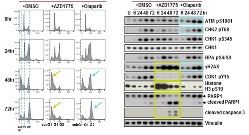
Figure 1(B): Olaparib and AZD1775 potentiate the efficacy of CRLX101 through different mechanisms. NCI-H417a cells were treated with CRLX101 for 48 hours either alone or in combination with indicated DDR inhibitors. Following washout of CRLX101, DMSO or DDR inhibitors were replenished. Samples were harvested at indicated timepoints for cell cycle status analysis by flow cytometry (left panel) or DDR biomarker activation analysis by immunoblotting (right panel).
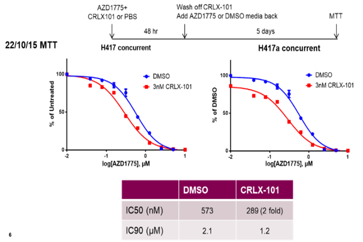
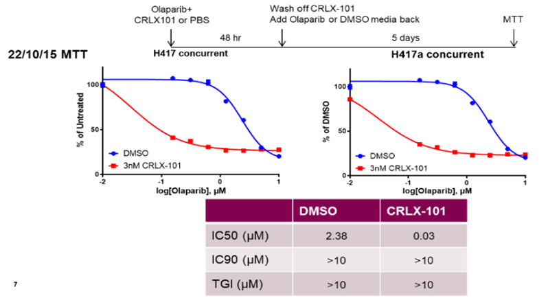
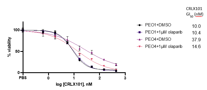
A significant proportion of cells (~20-30%) were not killed by CRLX101 treatment alone, even at the higher concentrations. Combinations with the PARPi, ATMi, or ATRi shifted the GI50 curve but did not result in any increase in this maximumcell kill value. Combination with the WEE1 inhibitor (AZD1775), although not significantly shifting the GI50 curve, did result in a more potent cell kill (>90 percentage cells). No obvious combination benefit was seen with DNA-PK inhibitor (AZ’6119) or the Aurora B kinase inhibitor (AZD1152). Combining a fixed dose of CRLX101 (3nM) with varying concentrations of olaparib or AZD1775 similarly demonstrated a shift in the efficacy curves of WEE1i and the PARPi. This effect was particularly striking for the olaparib dose response combination, where 0.3µM concentration of olaparib was sufficient to achieve greater than 50% growth inhibition (Supplementary Figure S1) suggesting that lower doses of olaparib could be used in vivo or in the clinical setting, in case there is a necessity to dose reduce due to normal tissue toxicities. Olaparib was also seen to lower the GI50 of CRLX101 by 2.6-fold in another cell line model, the ovarian cancer model POE4 (Supplemental Fig. S1).
Figure S1: CRLX101 potentiates the efficacy of WEE1 and PARP inhibitors in vitro
Figure S1: CRLX101 potentiates the efficacy of WEE1 and PARP inhibitors in vitro
Figure S1: CRLX101 potentiates the efficacy of WEE1 and PARP inhibitors in vitro
To understand the mechanism of action of different DDRi when combined with CRLX101, we initially compared combination treatments for CRLX101 with PARPi or WEE1i. First, we harvested NCI-H417 cells at various timepoints following treatment and analyzed cell cycle changes (Figure 1B, left) as well as the activation of DDR biomarkers (Figure 1B, right). CRLX101 activated a number of DDR markers, including pATM, pCHK2, pCHK1, pRPA and gH2AX. The treatment of CRLX101 (+DMSO control) resulted in a transient decrease in phospho-Histone H3 (pHH3), likely reflecting the induction of DNA damage (gH2AX) and replication stress (pS4/8 RPA) in S-phase. By contrast, the combination of CRLX101 with the WEE1 inhibitor AZD1775 leads to an initial increase in cells entering mitosis, since WEE1 inhibition, while increasing DNA damage and replication stress (gH2AX, pRPA and pCHK1), also overrides the G2/M checkpoint as indicated by pCDK1 inhibition and previously described (Young et al., 2019). In this study, following WEE1i treatment, an initial early mitotic entry of cancer cells that have already undergone DNA replication and were in late S or G2 phase occurred, followed by a reduction in mitotic entry as cells that had not undergone replication arrested in early S phase. In the NCI-H427a cells combination of CRLX101 with WEE1 inhibition therefore resulted in aberrant mitotic entry and enhanced cell death. The latter is indicated by increases in cleaved PARP and cleaved caspase 3 levels and extensive sub-G1 peak (Figure 1B). By contrast, the olaparib combination enhanced CRLX101-induced DNA damage, as indicated by elevated pATM and gH2AX, as well as activating the G2/M checkpoint (pCHK1 and pCDK1), with a concomitant reduction in pHH3. Combining PARP inhibitor with CRLX101 thus likely results in more DNA double-strand breaks and a prolonged cell cycle arrest in the late S and G2 phases (Figure 1B).
Increased hematological impact of concurrent CRXL101 and olaparib combination schedule
The strong mechanistic rationale for combining TOP1i with olaparib (Murai et al., 2014; Pommier et al., 2016) has previously encouraged the initiation of several Phase 1 clinical studies to investigate the tolerability of olaparib in combination with irinotecan (Chen et al., 2016) or topotecan (Samol et al., 2012). However, these combinations were not developed any further in the clinic, due to dose-limiting hematological adverse effects and a resulting sub-therapeutic combination maximum tolerated dose (MTD).
The pharmacokinetics and exposure levels of CRLX101 in both mouse and rats have previously been published and compared to exposures at different drug dose levels in humans that identified the human MTD (15mg/m2) equivalent dose for both species, namely 5mg/kg in the mouse and 2.5mg/kg in the rat. To assess CRLX-101 induced bone marrow toxicity in rats we used either 2.5mg/kg (MTD), 2mg/kg (80% MTD) or 1.25mg/kg (50% MTD), while for mouse xenograft efficacy studies, we used either 5mg/kg (MTD) or 4mg/kg (80% MTD).
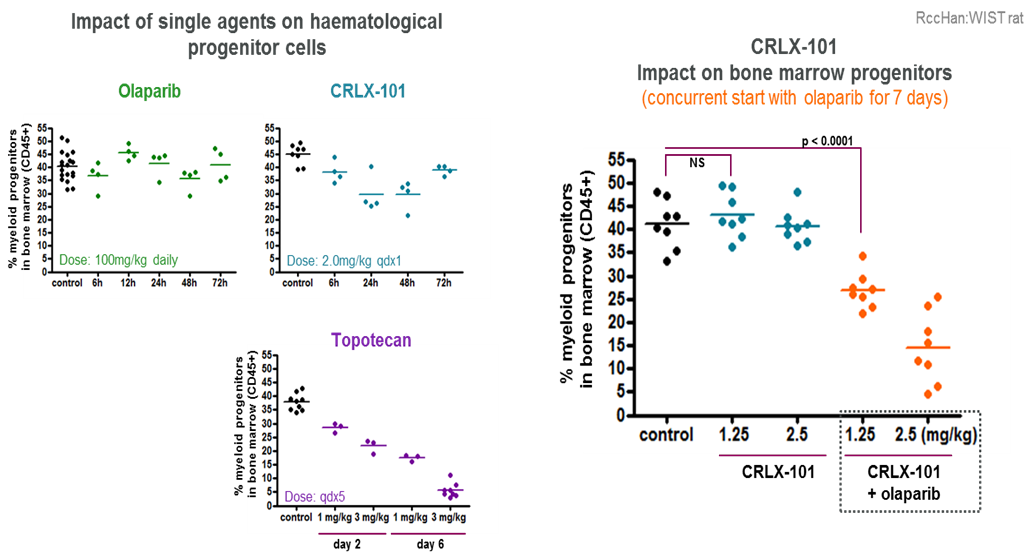
Assessment of bone marrow toxicity in immunocompetent RccHan:WIST rats treated with CRLX101 or olaparib as single agents, with topotecan included as a comparator, is shown in Figure 2A. At 48h, there was little effect of either olaparib or CRLX101 alone. By contrast, topotecan treatment produced a clear reduction in the myeloid progenitor (CD45+) population by day 2, with a nadir at day 6. However, the concurrent combination of olaparib and CRLX101 produced substantially greater bone marrow toxicity than either single agent alone. For the olaparib–CRLX101 combination, the nadir occurred at day 7, and the effects at this time point are shown in Figure 2B. In this rat bone marrow model, the combination effect was dose dependent and was observed when olaparib (100 mg/kg qdx7) was combined with CRLX101 at both the preclinical equivalent MTD and lower dose levels. Similar hematological effects of the combination were also observed in peripheral blood (Supplementary Figure S2).
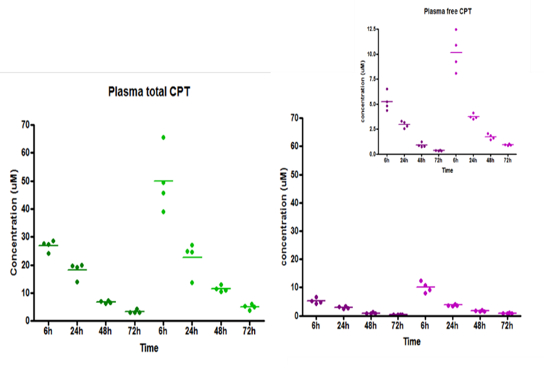
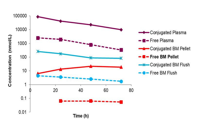
Figure S2: CRLX101 plasma and tumour pharmacokinetics
Figure S2: CRLX101 plasma and tumour pharmacokinetics
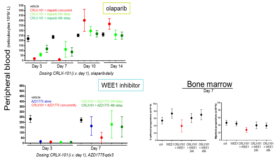
Assessment of the hematological effects of two other DDR inhibitors, namely the WEE1i (AZD1775) and the ATMi (AZD0156), was also carried out. WEE1i was chosen, since this gave the greatest maximum cell kill in combination with CRLX101 of all the DDRi tested, while ATMi was chosen because this gave the greatest level of potentiation (nearly 8-fold). Combinations of both agents demonstrated a greater degree of effect on the bone marrow than olaparib (Supplementary Figure S2).
CRXL101 treatment generates different DNA damage induction profiles in bone marrow vs. tumour
Our initial data from the rat bone marrow response studies (Fig. 2) indicated a potential risk that concurrent combination of CRLX101 with olaparib would lead to exacerbated hematological toxicity.
Figure 2: Increased hematological impact of CRLX101 in concurrent combination with olaparib. RccHan:WIST rats were treated with single agents or with concurrent CRLX101 plus olaparib; topotecan was included as a clinically relevant comparator. Bone marrow cells were harvested from the femur at the indicated time points and analysed by flow cytometry for frequencies of nucleated myeloid progenitor cells (LDS-751pos CD45pos).
Therefore, we sought to determine whether rational sequencing of the two agents could be explored to improve bone marrow tolerability, while not compromising on anti-tumour efficacy. To evaluate the effect of CRLX101 on DNA damage in bone marrow versus tumour cells, we treated preclinical models - rats for bone marrow analysis, and NCI-H417 tumour bearing nude mice – with equivalent, clinically relevant exposures of CRLX101 and harvested bone marrow and tumour tissue between 6h – 72h post dosing. Samples were processed for immunohistochemical staining with antibodies against γH2AX, as a marker of DNA double-strand breaks, or cleaved caspase 3, as a marker of apoptosis. Plasma, tumour, and bone marrow samples were also collected for pharmacokinetic analysis by mass spectrometry (Supplementary Figures S3 and S4).
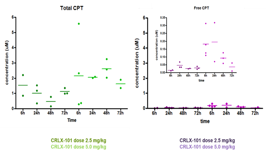
Figure S3:CRLX101 bone marrow pharmacokinetics
Figure S4:Hematological effects of CRLX101 combined with WEE1i or ATMi compared to olaparib
Figure S4:Hematological effects of CRLX101 combined with WEE1i or ATMi compared to olaparib
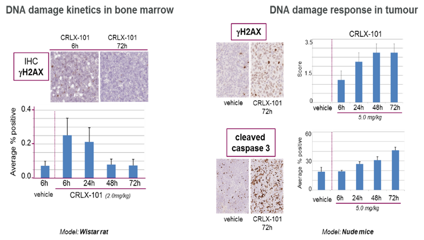
We found that only a small fraction of CRLX101 entered the bone marrow (<1% of plasma levels at 6–72h), resulting in low and transient DNA damage, as indicated by γH2AX between 6h and 24h, with little observed loss of bone marrow cellularity (Figure 3A). In contrast, CRLX101 is retained for a much longer time in tumour cells, leading to sustained DNA damage and associated cell death (Figure 3A).
Figure 3(A): CRLX101 exposure leads to distinct profiles of DNA damage induction in bone marrow versus tumour tissue. Animals received a clinically relevant dose of CRLX101, and tissues were harvested between 6h and 72h after dosing (bone marrow from wild-type rats; tumour from NCI-H417-bearing nude mice). Samples were analysed by immunohistochemistry for γH2AX, a marker of DNA double-strand breaks, or cleaved caspase 3, a marker of apoptosis.
A gap schedule of CRLX101 with olaparib reduces the impact on bone marrow
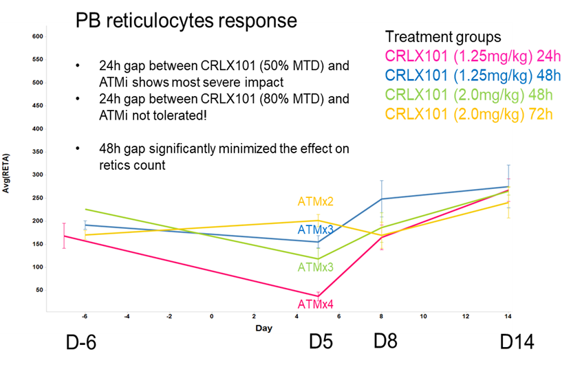
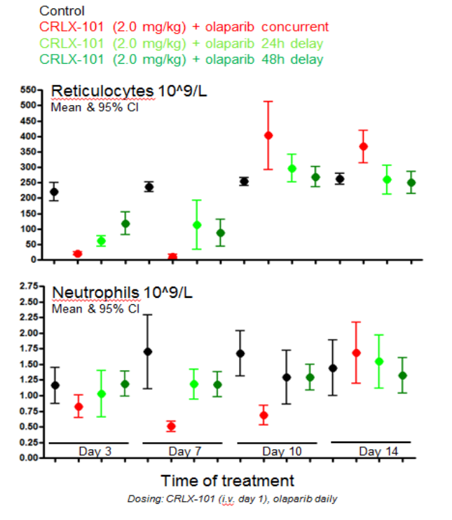
The pharmacodynamic biomarker analysis outlined in Figure 3A demonstrated clear differences following a single dose of CRLX101, between DNA damage induction and recovery in bone marrow vs. tumour tissue. (Figure 3A). Based on these results, we hypothesized that introducing at least a 24h gap between CRLX101 and olaparib dosing would allow bone marrow cells to recover from the initial CRLX101-induced damage before olaparib could potentiate this effect, thereby reducing the likelihood of cell death and facilitating hematological recovery. Given the dose-dependent effects seen in Figure 2, we chose an intermediate dose of CRLX101 for the gap schedule of 2mg/kg, or 80% of the equivalent clinical MTD. While 24h or 48h following CRLX101 treatment should provide the potential for bone marrow DNA damage resolution, the gH2AX biomarker data indicated this would not be the case for tumour cells since sustained and elevated levels of DNA damage, as well as the continued presence of the compound, were detected at 24h and at later time points. Thus, it would be predicted that even with a 24h or 48h gap, the effects of CRLX101 could be further potentiated by olaparib. To test this hypothesis, we treated rats with a single dose of CRLX101 and then dosed olaparib simultaneously or with a 24h or 48h delay. Peripheral blood was collected from tail veins and blood differentials analysed. Combination using a delayed or gap schedule of 24h or greater between the CRLX101 and the olaparib had a sparing effect on peripheral blood cells, in terms of both the extent of the nadir and the time to recovery (Figure 3B).
Figure 3(B):Sequenced (“gap”) schedules of CRLX101 and olaparib reduce hematological impact. Wistar rats received a single dose of CRLX101, followed by olaparib administered either concurrently or after a 24h or 48h delay. Peripheral blood was collected from the tail vein and analysed on the Siemens Advia 2120i haematology analyser.
A sequenced (gap) schedule of CRLX101 with olaparib maintains anti-tumour efficacy
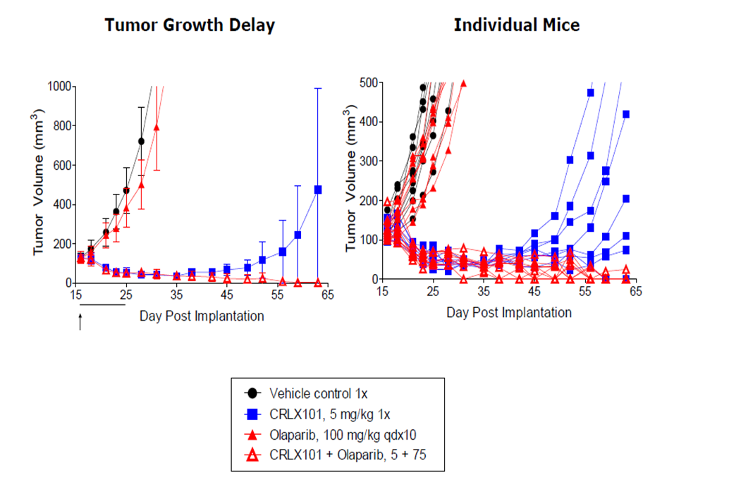
A previous SCLC in vivo study of concurrent treatment of olaparib with CRLX101 in the xenograft model NCI-H1048 demonstrated that the combination anti-tumour activity was more effective than CRLX101 treatment alone, with 0/7 tumour progressions in the combination cohort versus 7/7 for CRLX101 as a single agent (Supplementary Figure S5).
Figure S5:Enhanced activity of CRLX101 when in combination with olaparib in the SCLC xenograft in vivo model NCI-H480
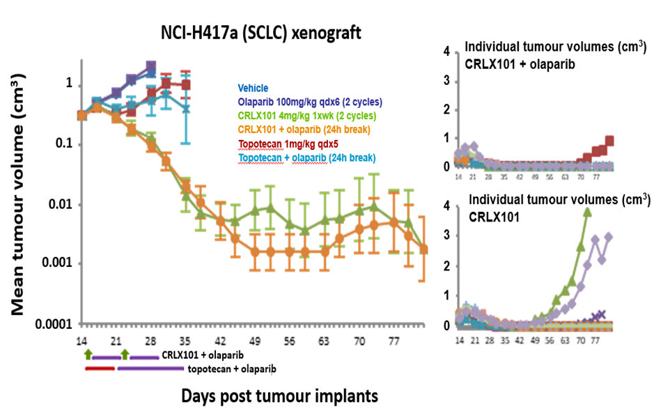
To test whether gap scheduling of CRLX101 and olaparib can maintain enhanced anti-tumour efficacy of the combination over single agent CRLX101 alone, we tested the response in vivo in a small cell lung cancer NCI-H417 subcutaneously implanted xenograft model using 4mg/kg CRLX101 (one dose/week), which is 80% of the clinical MTD equivalent in mice, with daily dosing of 100mg/kg olaparib (MTD equivalent) following a 24h gap for two weeks. As a comparator, we used the 4mg/kg single agent dose of CRLX101, single agent olaparib, and topotecan at the MTD for this agent of 1mg/kg for five days, both as a single agent and in combination with olaparib in a 24h gap schedule. Individual tumour volumes were measured regularly up to 83 days. This study demonstrated that CRLX101 in combination with olaparib was more effective than CRLX101 alone and was significantly more efficacious than topotecan administered at its MTD (Figure 4A).
Figure 4(A): Gap scheduling of olaparib following CRLX101 treatment maintains anti-tumour efficacy compared to single agent CRLX101 alone. Human small cell lung cancer cells (NCI-H417a) were implanted subcutaneously in athymic Fox1-nu mice. Animals were dosed with individual agents or combination of CRLX-101 (1xwk) + olaparib (qdx10, 24h after) or topotecan (qdx5) + olaparib (qdx14, 24h after). Individual tumour volumes were measured regularly up to 83 days.
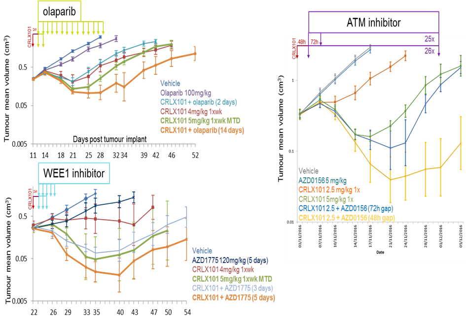
Given that the in vivo model we were using did not have an identified homologous recombination repair (HRR) deficiency that would provide olaparib single-agent sensitivity, we asked the question of whether there would be any benefit in extending the dosing of olaparib beyond a couple of days after CRLX101 treatment. Mice implanted with NCI-H417a tumour cells were therefore treated with a single dose of CRLX101 (4mg/kg) and after 24h, olaparib (100mg/kg) for either 2 days or 14 days. The data depicted in Figure 4B clearly demonstrate that dosing olaparib for 14 days is significantly better than for just 2 days, even in a homologous recombination repair-proficient background. The most likely explanation for the enhanced potentiation provided by extended olaparib treatment is that CRLX101 is retained in tumours and continues to generate DNA damage over time, the effects of which are enhanced by the continued presence of olaparib.
Finally, to extend our in vitro data suggesting that the WEE1i and ATMi may also potentiate the activity of CRLX101, we further tested these two combinations in the NCI-H417a tumour xenograft model (Figure 4B).
Figure 4(B): Extending the duration of olaparib treatment following CRLX101 treatment maximises the anti-tumour effect of the combination. Mice implanted with NCI-H417a tumour cells were dosed with single dose of CRLX101 + olaparib (100 m/kg) for 2 days or 14 days (top panel) or CRLX101 + AZD1775 (120mg/kg) for 3 or 5 days (bottom panel) starting 24h after the CRLX101 or CRXL101 + AZD0156 (5mg/kg) for up to 26 days.
Mice were treated with CRLX101 (4mg/kg) plus AZD1775 (120mg/kg) for 3 or 5 days starting 24h after the CRLX101. The combination of reduced dose of CRLX101 (4mg/kg) with 5 days of AZD1775 dosing led to better efficacy than the 3-day schedule and was also more potent than CRLX101 monotherapy at its maximum tolerated dose (5mg/kg). However, clinical data on adavosertib/AZD1775 suggests it will not be possible to be able to deliver the 120mg/kg equivalent dose in humans for five days (Do et al., 2015). For the ATMi, initial tolerability studies in rats showed that a combination with CRLX101 dosed 24h apart was not tolerated. Reflecting on this observation and the fact that the ATMi was the most potent potentiator of CRLX101 activity in vitro (Figure 1), we focused on 48h and 72h gap schedules and further reduced the dose of CRLX101 to 50% MTD equivalent (2.5mg/kg in mice) for this combination. It was encouraging to see that the combination of 2.5mg/kg of CRLX101 with AZD0156 dosed 48h apart led to tumour regression, significantly greater than full dose of CRXL101 alone (Figure 4B). However, bone marrow tolerability would be a potential concern. The anti-tumour efficacy was still maintained with the 72h gap schedule; however, the effect was just marginally greater than CRLX101 alone when dosed in its MTD.
Taken together, our data suggested that while there were a number of options for DDRi combinations with CRLX101, the most likely to succeed clinically was the PARPi olaparib combination. The preclinical studies suggested a combination dose and schedule of 80% of the clinical MTD equivalent of CRLX101 followed by a 48h gap and then daily olaparib using a dose escalation approach to get as close to the MTD as possible until two days before the next CRLX101 treatment begins, to avoid concurrent dosing at the beginning of the second cycle. Based on these insights from preclinical studies, a phase I clinical trial combining olaparib and CRLX101 has been designed and carried out to assess the gap schedule approach (NCT02769962).
DISCUSSION
Many SoC chemotherapies act by generating DNA damage that has the potential to be enhanced by inhibitors of the DDR. However, overlapping toxicities, in particular bone marrow toxicity, has hampered the ability to combine these agents. Attempting to identify an effective and sufficiently well tolerated dose and schedule in the clinic cantake many years and still fail to identify a combination that is clearly better than the full dose SoC chemotherapy alone. A good illustration of this is the combination of the PARPi olaparib with carboplatin and paclitaxel, where a Phase I trial (NCT00516724) began in 2007, took many years to complete and involved 189 patients. Only recently has this trial been reported (van der Noll et al., 2020) and the eventual conclusion was that olaparib in combination with carboplatin and/or paclitaxel resulted in increased hematologic toxicities, making it challenging to establish a dosing regimen that could be tolerated for multiple cycles without dose modifications. Anotherexample, where there is a clear mechanistic rational for combination but where enhanced bone marrow toxicity has meant no optimal recommended Phase II dose has been identified that could be taken forward, is the combination of PARPi with TOP1i such as irinotecan and topotecan (Murai et al., 2014). Several clinical trials (Chen et al., 2016; Dhawan et al., 2017; Hendrickson et al., 2018; Kummar et al., 2011; LoRusso et al., 2016; Rajan et al., 2012; Samol et al., 2012) have tried and failedto combine PARPiand TOP1i at doses close to their individual MTDs with dose limiting myelosuppression precluding administration of >20%of a PARPi MTD dose.
One limitation of the work presented here is that the experiments were performed in selected tumour models, and the magnitude of benefit may vary according to tumour biology, including DNA repair status and intrinsic sensitivity to TOP1 inhibition. Moreover, the feasibility of this approach is likely to depend on the pharmacokinetic and pharmacodynamic properties of the DNA-damaging partner, particularly the extent to which sustained tumour retention can be separated from normal tissue exposure.
Nonetheless, the data presented in this preclinical study deploying a gap schedule that could deliver 80% of the MTD equivalent of CRLX101 with an MTD equivalent of olaparib, was used to design a clinical trial (Clinical trials.gov reference NCT02769962, where CRLX101 was renamed EP0057). This clinical study has recently been published and provides encouraging validation of our gap scheduling approach, with escalation of both the PARP inhibitor and TOP1 inhibitor to approximately 80% of their respective single-agent MTDs, consistent with our preclinical data, and higher than previous clinical trials were able to achieve and with encouraging clinical activity in heavily pretreated patients (Thomas et al, 2025).
In addition to the specificexample of CRLX101that was the focus of this study, the gap scheduling approach exemplified here also has the potential to work for PARPi with other targeted DNA damaging agents and for other DDRi. One exciting opportunity will be the combination of DDRi with antibody drug conjugates (ADCs) and targeted radioligands that could benefit from gap combination schedules with DDR inhibitors (Yap et al., 2026).
CONCLUSION
In summary, DDR inhibitor combinations, either with DNA damaging agents or other DDR inhibitors, have significant therapeutic potential but are nevertheless challenging. The work presented here has highlighted how an assessment of preclinical models of both tumour and normal tissue responses, such as those of rat bone marrow, can provide dose and scheduling insights that can significantly enable combination testing in the clinic.
Acknowledgements
We would like to thank those currentand past AstraZeneca scientists, who are not co-authors but who nevertheless contributed towards the work, namely Jamie Reens, Catherine Wilkinson, Anna Cronin, Richard Knights and Pete Newham.
Author Contributions
Original conceptualization and methodology for the preclinical studies was provided by MOC and LOC. Experimental investigation was provided by LOC, AW, CS, JB, RO, AS, GH, JR, AL and EC, with overall supervision from MOC.
Declaration of interests
MOC, LOC, AW, AS, GH, AL and EC are employees and shareholders of AstraZeneca, while CS, JB, and RO are former employees of AstraZeneca.
References
Angius, G., Tomao, S., Stati, V., Vici, P., Bianco, V., and Tomao, F. (2020). Prexasertib, a checkpoint kinase inhibitor: from preclinical data to clinical development. Cancer Chemoth Pharm 85, 9-20. View
at PublisherView
at Google Scholar
Ashton, S., Song, Y. H., Nolan, J., Cadogan, E., Murray, J., Odedra, R., Foster, J., Hall, P. A., Low, S., Taylor, P., et al. (2016). Aurora kinase inhibitor nanoparticles target tumors with favorable therapeutic index in vivo. Sci Transl Med 8. View
at PublisherView
at Google Scholar
Beck, A., Goetsch, L., Dumontet, C., and Corvaia, N. (2017). Strategies and challenges for the next generation of antibody drug conjugates. Nat Rev Drug Discov 16, 315-337. View
at PublisherView
at Google Scholar
Chen, E. X., Jonker, D. J., Siu, L. L., McKeever, K., Keller, D., Wells, J., Hagerman, L., and Seymour, L. (2016). A Phase I study of olaparib and irinotecan in patients with colorectal cancer: Canadian Cancer Trials Group IND 187. Investigational new drugs 34, 450-457. View
at PublisherView
at Google Scholar
Cheung-Ong, K., Giaever, G., and Nislow, C. (2013). DNAdamaging agents in cancer chemotherapy: serendipity and chemical biology. Chem Biol 20, 648-659. View
at PublisherView
at Google Scholar
Ciccia, A., and Elledge, S. J. (2010). The DNA damage response: making it safe to play with knives. Mol Cell 40, 179-204. View
at PublisherView
at Google Scholar
Clark, A. J., Wiley, D. T., Zuckerman, J. E., Webster, P., Chao, J., Lin, J., Yen, Y., and Davis, M. E. (2016). CRLX101 nanoparticles localize in human tumors and not in adjacent, nonneoplastic tissue after intravenous dosing. Proc Natl Acad Sci U S A 113, 3850-3854. View
at PublisherView
at Google Scholar
Coleman, R. L., Oza, A. M., Lorusso, D., Aghajanian, C., Oaknin, A., Dean, A.,): a randomised, double-blind, placebo-controlled, phase 3 trial. Lancet 390, 1949- 1961. View
at PublisherView
at Google Scholar
Coussy, F., El-Botty, R., Chateau-Joubert, S., Dahmani, A., Montaudon, E., Leboucher, S., Morisset, L., Painsec, P., Sourd, L., Huguet, L., et al. (2020). BRCAness, SLFN11, and RB1 loss predict response to topoisomerase I inhibitors in triple-negative breast cancers. Sci Transl Med 12. View
at PublisherView
at Google Scholar
Curtin, N. J. (2012). DNA repair dysregulation from cancer driver to therapeutic target. Nat Rev Cancer 12, 801-817. View
at PublisherView
at Google Scholar
de Bono, J., Mateo, J., Fizazi, K., Saad, F., Shore, N., Sandhu, S., Chi, K. N., Sartor, O., Agarwal, N., Olmos, D., et al. (2020). Olaparib for Metastatic Castration-Resistant Prostate Cancer. N Engl J Med 382, 2091-2102. View
at PublisherView
at Google Scholar
Do, K., Wilsker, D., Ji, J., Zlott, J., Freshwater, T., Kinders, R. J., Collins, J., Chen, A. P., Doroshow, J. H., and Kummar, S. (2015). Phase I Study of Single-Agent AZD1775 (MK- 1775), a Wee1 Kinase Inhibitor, in Patients With Refractory Solid Tumors. J Clin Oncol 33, 3409-3415. View
at PublisherView
at Google Scholar
Durant, S. T., Zheng, L., Wang, Y. C., Chen, K., Zhang, L. L., Zhang, T. W., Yang, Z. F., Riches, L., Trinidad, A. G., Fok, J. H. L., et al. (2018). The brain-penetrant clinical ATM inhibitor AZD1390 radiosensitizes and improves survival of preclinical brain tumor models. Sci Adv 4. View
at PublisherView
at Google Scholar
Fok, J. H. L., Ramos-Montoya, A., Vazquez-Chantada, M., Wijnhoven, P. W. G., Follia, V., James, N., Farrington, P. M., Karmokar, A., Willis, S. E., Cairns, J., et al. (2019). AZD7648 is a potent and selective DNA-PK inhibitor that enhances radiation, chemotherapy and olaparib activity. Nat Commun 10. View
at PublisherView
at Google Scholar
Fong, P. C., Boss, D. S., Yap, T. A., Tutt, A., Wu, P., Mergui- Roelvink, M., Mortimer, P., Swaisland, H., Lau, A., O'Connor, M. J., et al. (2009). Inhibition of poly(ADPribose) polymerase in tumors from BRCA mutation carriers. N Engl J Med 361, 123-134. View
at PublisherView
at Google Scholar
Foote, K. M., Nissink, J. W. M., McGuire, T., Turner, P., Guichard, S., Yates, J. W. T., Lau, A., Blades, K., Heathcote, D., Odedra, R., et al. (2018). Discovery and Characterization of AZD6738, a Potent Inhibitor of Ataxia Telangiectasia Mutated and Rad3 Related (ATR) Kinase with Application as an Anticancer Agent. Journal of Medicinal Chemistry 61, 9889-9907. View
at PublisherView
at Google Scholar
Golan, T., Hammel, P., Reni, M., Van Cutsem, E., Macarulla, T., Hall, M. J., Park, J. O., Hendrickson, A. E. W., Menefee, M. E., Hartmann, L. C., Long, H. J., Northfelt, D. W., Reid, J. M., Boakye-Agyeman, F., Kayode, O., Flatten, K. S., Harrell, M. I., et al. (2018). A Phase I Clinical Trial of the Poly(ADP-ribose) Polymerase Inhibitor Veliparib and Weekly Topotecan in Patients with Solid Tumors. Clinical Cancer Research 24, 744-752. View
at PublisherView
at Google Scholar
Hirai, H., Iwasawa, Y., Okada, M., Arai, T., Nishibata, T., Kobayashi, M., Kimura, T.,Jackson, S. P., and Bartek, J. (2009). The DNA-damage response in human biology and disease. Nature 461, 1071-1078. View
at PublisherView
at Google Scholar
Kummar, S., Chen, A., Ji, J., Zhang, Y., Reid, J. M., Ames, M., Jia, L., Weil, M., Speranza, G., Murgo, A. J., et al. (2011). Phase I study of PARP inhibitor ABT-888 in combination with topotecan in adults with refractory solid tumors and lymphomas. Cancer research 71, 5626-5634. View
at PublisherView
at Google Scholar
Ledermann, J., Harter, P., Gourley, C., Friedlander, M., Vergote, I., Rustin, G., Scott, C. L., Meier, W., Shapira-Frommer, R., Safra, T., et al. (2014). Olaparib maintenance therapy in patients with platinum-sensitive relapsed serous ovarian cancer: a preplanned retrospective analysis of outcomes by BRCA status in a randomised phase 2 trial. Lancet Oncol 15, 852-861. View
at PublisherView
at Google Scholar
Litton, J. K., Rugo, H. S., Ettl, J., Hurvitz, S. A., Goncalves, A., Lee, K. H., Fehrenbacher, L., Yerushalmi, R., Mina, L. A., Martin, M., et al. (2018). Talazoparib in Patients with Advanced Breast Cancer and a Germline BRCA Mutation. N Engl J Med 379, 753-763. View
at PublisherView
at Google Scholar
LoRusso, P. M., Li, J., Burger, A., Heilbrun, L. K., Sausville, E. A., Boerner, S. A., Smith, D., Pilat, M. J., Zhang, J., Tolaney, S. M., et al. (2016). Phase I Safety, Pharmacokinetic, and Pharmacodynamic Study of the Poly(ADP-ribose) Polymerase (PARP) Inhibitor Veliparib (ABT-888) in Combination with Irinotecan in Patients with Advanced Solid Tumors. Clinical cancer research : an official journal of the American Association for Cancer Research 22, 3227-3237. View
at PublisherView
at Google Scholar
Menear, K. A., Adcock, C., Boulter, R., Cockcroft, X. L., Copsey, L., Cranston, A., Dillon, K. J., Drzewiecki, J., Garman, S., Gomez, S., et al. (2008). 4-[3-(4-cyclopropanecarbonylpiperazine-1-carbonyl)-4-fluorobenzyl]-2H-phthalazin- 1-one: a novel bioavailable inhibitor of poly(ADP-ribose) polymerase-1. J Med Chem 51, 6581-6591. View
at PublisherView
at Google Scholar
Min, A., Im, S. A., Jang, H., Kim, S., Lee, M., Kim, D. K., Yang, Y., Kim, H. J., Lee, K. H., Kim, J. W., et al. (2017). AZD6738, A Novel Oral Inhibitor of ATR, Induces Synthetic Lethality with ATM Deficiency in Gastric Cancer Cells. Molecular Cancer Therapeutics 16, 566-577. View
at PublisherView
at Google Scholar
Mirza, M. R., Monk, B. J., Herrstedt, J., Oza, A. M., Mahner, S., Redondo, A., Fabbro, M., Ledermann, J. A., Lorusso, D., Vergote, I., et al. (2016). Niraparib Maintenance Therapy in Platinum-Sensitive, Recurrent Ovarian Cancer. N Engl J Med 375, 2154-2164. View
at PublisherView
at Google Scholar
Murai, J., Zhang, Y., Morris, J., Ji, J., Takeda, S., Doroshow, J. H., and Pommier, Y. G. (2014). Rationale for PARP inhibitors in combination therapy with camptothecins or temozolomide based on PARP trapping versus catalytic inhibition. The Journal of pharmacology and experimental therapeutics 349, 408-416. View
at PublisherView
at Google Scholar
'Connor, L. O., Rulten, S. L., Cranston, A. N., Odedra, R., Brown, H., Jaspers, J. E., Jones, L., Knights, C., Evers, B., Ting, A., et al. (2016). The PARP Inhibitor AZD2461 Provides Insights into the Role of PARP3 Inhibition for Both Synthetic Lethality and Tolerability with Chemotherapy in Preclinical Models. Cancer Research 76, 6084-6094. View
at PublisherView
at Google Scholar
Pham, E., Birrer, M. J., Eliasof, S., Garmey, E. G., Lazarus, D., Lee, C. R., Man, S., Matulonis, U. A., Peters, C. G., Xu, P., et al. (2015). Translational Impact of Nanoparticle-Drug Conjugate CRLX101 with or without Bevacizumab in Advanced Ovarian Cancer. Clinical Cancer Research 21, 808-818. View
at PublisherView
at Google Scholar
Pilie, P. G., Gay, C. M., Byers, L. A., O'Connor, M. J., and Yap, T. A. (2019). PARP Inhibitors: Extending Benefit Beyond BRCA-Mutant Cancers. Clin Cancer Res 25, 3759-3771. View
at PublisherView
at Google Scholar
Pommier, Y., O'Connor, M. J., and de Bono, J. (2016). Laying a trap to kill cancer cells: PARP inhibitors and their mechanisms of action. Sci Transl Med 8, 362ps317 View
at PublisherView
at Google Scholar
Rajan, A., Carter, C. A., Kelly, R. J., Gutierrez, M., Kummar, S., Szabo, E., Yancey, M. A., Ji, J. P., Mannargudi, B., Woo, S., et al. (2012). A Phase I Combination Study of Olaparib with Cisplatin and Gemcitabine in Adults with Solid Tumors. Clinical Cancer Research 18, 2344-2351. View
at PublisherView
at Google Scholar
Reaper, P. M., Griffiths, M. R., Long, J. M., Charrier, J. D., MacCormick, S., Charlton, P. A., Golec, J. M. C., and Pollard, J. R. (2011). Selective killing of ATM- or p53-deficient cancer cells through inhibition of ATR. Nat Chem Biol 7, 428-430. View
at PublisherView
at Google Scholar
Riches, L. C., Trinidad, A. G., Hughes, G., Jones, G. N., Hughes, A. M., Thomason, A. G., Gavine, P., Cui, A., Ling, S., Stott, J., et al. (2020). Pharmacology of the ATM Inhibitor AZD0156: Potentiation of Irradiation and Olaparib Responses Preclinically. Molecular Cancer Therapeutics 19, 13-25. View
at PublisherView
at Google Scholar
Robson, M., Im, S. A., Senkus, E., Xu, B., Domchek, S. M., Masuda, N., Delaloge, S., Li, W., Tung, N., Armstrong, A., et al. (2017). Olaparib for Metastatic Breast Cancer in Patients with a Germline BRCA Mutation. N Engl J Med 377, 523-533. View
at PublisherView
at Google Scholar
Samol, J., Ranson, M., Scott, E., Macpherson, E., Carmichael, J., Thomas, A., and Cassidy, J. (2012). Safety and tolerability of the poly(ADP-ribose) polymerase (PARP) inhibitor, olaparib (AZD2281) in combination with topotecan for the treatment of patients with advanced solid tumors: a phase I study. Invest New Drug 30, 1493-1500. View
at PublisherView
at Google Scholar
Thomas, A., Teicher, B. A., and Hassan, R. (2016). Antibody-drug conjugates for cancer therapy. Lancet Oncol 17, e254-e262. View
at PublisherView
at Google Scholar
Thomas, A., Takahashi, N., Oplustil O'Connor, L., Redon, C.E., Mohindroo, C., Sciuto, L., Pongor, L., Schmidt, K.T., Steinberg, S.M., Aladjem, M.I., Figg, W.D., O'Connor, M. J., Pommier, Y. (2025). Tumor-targeted top1 inhibitor delivery with optimized parp inhibition in advanced solid tumors: a phase i trial of gapped scheduling. Nat. Comm. 16, View
at PublisherView
at Google Scholar
van der Noll, R., Jager, A., Ang, J. E., Marchetti, S., Mergui-Roelvink, M. W. J., de Bono, J. S., Lolkema, M. P., de Jonge, M. J. A., van der Biessen, D. A., Brunetto, A. T., et al. (2020). Phase I study of intermittent olaparib capsule or tablet dosing in combination with carboplatin and paclitaxel (part 2). Invest New Drugs 38, 1096-1107. View
at PublisherView
at Google Scholar
Wang, A. Z., Langer, R., and Farokhzad, O. C. (2012). Nanoparticle Delivery of Cancer Drugs. Annu Rev Med 63, 185-198. View
at PublisherView
at Google Scholar
Weiss, G. J., Chao, J., Neidhart, J. D., Ramanathan, R. K., Bassett, D., Neidhart, J. A., Choi, C. H. J., Chow, W., Chung, V., Forman, S. J., et al. (2013). First-in-human phase 1/2a trial of CRLX101, a cyclodextrin-containing polymer-camptothecin nanopharmaceutical in patients with advanced solid tumor malignancies. Invest New Drugs 31, 986-1000. View
at PublisherView
at Google Scholar
Wengner, A. M., Siemeister, G., Lucking, U., Lefranc, J., Wortmann, L., Lienau, P., Bader, B., Bomer, U., Moosmayer, D., Eberspacher, U., et al. (2020). The Novel ATR Inhibitor BAY 1895344 Is Efficacious as Monotherapy and Combined with DNA Damage-Inducing or Repair-Compromising Therapies in Lenka Oplustil O’Connor, Anderson Wang, Claire Sadler, Jennifer Barnes, Rajesh Odedra, Aaron Smith, Gareth Hughes, Alan Lau, Andres Tellez, Scott Eliasof, Elaine Cadogan, Mark J. O’Connor (2026), Gap Scheduling of a PARP Inhibitor and Nanoparticle TOP1 Agent Combination Avoids Synergistic Bone Marrow Toxicity; J. Clinical Cancer and Oncology, 3(4): DOI: 10.64585/3065-3320-3-4-09. Mark J. O’Connor 22 | Clinical Cancer and Oncology Preclinical Cancer Models. Molecular Cancer Therapeutics 19, 26-38. View
at PublisherView
at Google Scholar
Wilkinson, R. W., Odedra, R., Heaton, S. P., Wedge, S. R., Keen, N. J., Crafter, C., Foster, J. R., Brady, M. C., Bigley, A., Brown, E., et al. (2007). AZD1152, a selective inhibitor of Aurora B kinase, inhibits human tumor xenograft growth by inducing apoptosis. Clinical Cancer Research 13, 3682-3688. View
at PublisherView
at Google Scholar
Wise, H. C., Iyer, G. V., Moore, K., Temkin, S. M., Gordon, S., Aghajanian, C., and Grisham, R. N. (2019). Activity of M3814, an Oral DNA-PK Inhibitor, In Combination with Topoisomerase II Inhibitors in Ovarian Cancer Models. Sci Rep-Uk 9. View
at PublisherView
at Google Scholar
Yap, T.A., Manning, H.C., Sapra, P., Mills, G.B., O'Connor, M.J. (2026). Targeting genomic instability in cancer. Cell 189, 2278-2306. View
at PublisherView
at Google Scholar
Young, L. A., O'Connor, L. O., de Renty, C., Veldman-Jones, M. H., Dorval, T., Wilson, Z., Jones, D. R., Lawson, D., Odedra, R., Maya-Mendoza, A., et al. (2019). Differential Activity of ATR and WEE1 Inhibitors in a Highly Sensitive Subpopulation of DLBCL Linked to Replication Stress. Cancer Res 79, 3762-3775. View
at PublisherView
at Google Scholar
"ScienceHood Publishing exceeded our expectations with their seamless execution and professionalism. Their team ensured timely communication, high-quality production, and attention to detail throughout the process. They transformed our vision into reality, delivering exceptional results. We highly recommend them for their efficiency, expertise, and commitment to excellence in publishing."
Lara Simmons
"ScienceHood Publishing surpassed our expectations with their professionalism, timely communication, and exceptional attention to detail. They transformed our vision into reality with outstanding results. We highly recommend them for their expertise and commitment to excellence."
Shippora Smith
"Publishing in this journal gave me the opportunity to involve Bioinformatics, which is a cutting-edge field with so much potential, into my previous research on Optogenetics and Artificial Intelligence. This experience helped me better understand how closely interconnected STEM fields truly are, and how many opportunities exist for interdisciplinary collaboration to allow them to work together and, once again, change the world, as science has always done"
Tomas Hadi Acosta Aguilera
"Dear editors,
It has also been a real pleasure working with you on the publication of our article. Your patience, understanding, and prompt responses whenever we needed them have been a great help to us in this joint endeavour."


 :
2026 Mark J. O’Connor. This open-access article is distributed under the terms of The Creative Commons Attribution License, which permits unrestricted use, distribution, and reproduction in any medium, provided the original author and source are credited.
:
2026 Mark J. O’Connor. This open-access article is distributed under the terms of The Creative Commons Attribution License, which permits unrestricted use, distribution, and reproduction in any medium, provided the original author and source are credited.